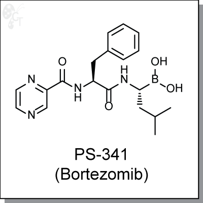
PS-341 (Bortezomib) | Proteasome inhibitorCellagen TechnologyPrice from $89.00 to $712.00.
Cellagen Technology
Proteasome inhibitor
MG-132 is a triterpene, peptide-aldehyde proteasome inhibitor derived from a Chinese medicinal plant. It inhibits 20S proteasome activty by covalently binding to the active site of the beta subunits and effectively blocks the proteolytic activity of the 26S proteasome complex. Through the formation of reactive oxygen species, MG-132 inhibits tumor cell growth by inducing cell cycle arrest and triggering apoptosis. [1]
MG-132 was found to enhance IL-6 expression in HUVEC. MG-132 does not affect IkB levels, suggesting that proteasome inhibition does not influence the NF-kB system without any agonist stimulation. In similar studies, MG-132 is believed to inhibit the degradation of phosphorylated MEK1/2 and activate the downstream region of this signalling cascade. [2]
In NCIH2452 and NCI H2052 human thoracic malignant pleural mesothelioma (MPM) cell lines, it was foud that 0.5 uM concentrations of MG-132 caused significant apoptosis as evidenced by DNA damage, cleavage of PARP and caspases 3, 7, and 9. [3]

Cellagen Technology
Proteasome inhibitor
MG-132 is a triterpene, peptide-aldehyde proteasome inhibitor derived from a Chinese medicinal plant. It inhibits 20S proteasome activty by covalently binding to the active site of the beta subunits and effectively blocks the proteolytic activity of the 26S proteasome complex. Through the formation of reactive oxygen species, MG-132 inhibits tumor cell growth by inducing cell cycle arrest and triggering apoptosis. [1]
MG-132 was found to enhance IL-6 expression in HUVEC. MG-132 does not affect IkB levels, suggesting that proteasome inhibition does not influence the NF-kB system without any agonist stimulation. In similar studies, MG-132 is believed to inhibit the degradation of phosphorylated MEK1/2 and activate the downstream region of this signalling cascade. [2]
In NCIH2452 and NCI H2052 human thoracic malignant pleural mesothelioma (MPM) cell lines, it was foud that 0.5 uM concentrations of MG-132 caused significant apoptosis as evidenced by DNA damage, cleavage of PARP and caspases 3, 7, and 9. [3]